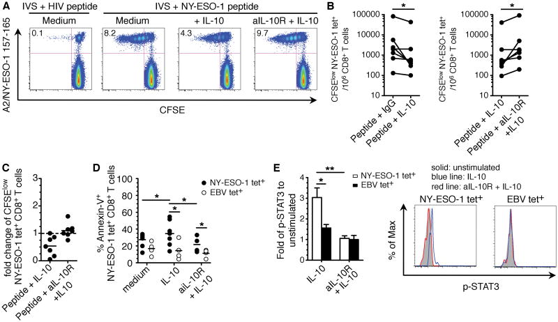
Figure 4.
IL-10 inhibits NY-ESO-1–specific CD8+ T cell proliferation after prolonged antigen stimulation. A and B, Dot plots from a representative patient (A) and summary for seven patients with melanoma (B) showing the variation in the frequencies of CFSElo NY-ESO-1–specific CD8+ T cells for 106 CD8+ T cells. CFSE-labeled PBMCs from patients were incubated with NY-ESO-1 157–165 peptide and rIL-10 or blocking mAb against IL-10R (aIL-10R) plus rhIL-10 or an isotype control antibody (IgG) before the evaluation of A2/NY-ESO-1 157–165 tet+ CD8+ T cell proliferation by flow cytometry. C, Fold change of the frequencies of CFSElo NY-ESO-1–specific CD8+ T cells after IVS with cognate peptide and rhIL-10 or aIL-10R plus rhIL-10. The ratios of the percentages of CFSElo NY-ESO-1–specific CD8+ T cells in the presence of rhIL-10 or in the presence of aIL-10R plus rhIL-10 and IgG isotype control are shown. D, Pooled data showing the percentage of Annexin-V+ determined by flow cytometry for A2/NY-ESO-1 157–165 tet+ CD8+ T cells (n = 7) or A2/EBV BMLF1 280–288 tet+ CD8+ T cells (n = 4), which were incubated for 6 d with NY-ESO-1 157–165 peptide or BMLF1 280–288 peptide and rhIL-10 or aIL-10R plus rhIL-10 or IgG. E, p-STAT3 expression measured by flow cytometry in A2/NY-ESO-1 157–165 tet+ CD8+ T cells or A2/EBV BMLF1 280–288 tet+ CD8+ T cells from melanoma patients (n = 5) treated or not with aIL-10R and then stimulated or not with IL-10.

