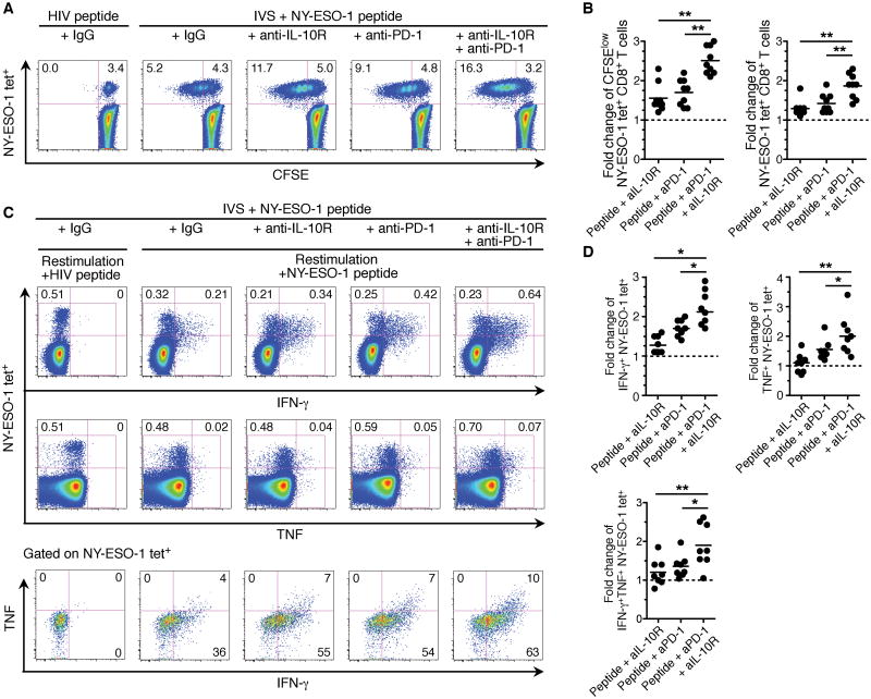
Figure 5.
IL-10R blockade adds to PD-1 blockade to increase the expansion and functions of NY-ESO-1–specific CD8+ T cells. A, Representative flow cytometry analysis from one melanoma patient showing the percentages of CFSElo NY-ESO-1–specific CD8+ T cells among total CD8+ T cells in CFSE-based proliferation assay (n = 9). B, Fold change of the frequencies of CFSElo (left) and total (right) NY-ESO-1–specific CD8+ T cells after IVS with cognate peptide and aPD-1 and/or aIL-10R. The ratio of the percentages of CFSElo and total NY-ESO-1–specific CD8+ T cells in the presence of indicated antibody treatment and isotype control antibody is shown. C, Representative flow cytometry analysis from one melanoma patient showing the percentages of IFN-γ- and TNF-producing NY0-ESO-1–specific CD8+ T cells among total CD8+ T cells (upper) and the percentages of IFN-γ+TNF+ NY-ESO-1–specific CD8+ T cells among NY-ESO-1–specific CD8+ T cells (lower). PBMCs were incubated with NY-ESO-1 157–165 peptide or with HIVpol 476–484 peptide and aPD-1 and/or aIL-10R or an isotype control antibody (IgG), prior to evaluating intracellular cytokine production of NY-ESO-1–specific CD8+ T cells upon stimulation with cognate peptide, (n=8). D, Fold change of the frequencies of IFN-γ +, TNF+ and IFN-γ+TNF+ NY-ESO-1–specific CD8+ T cells after IVS with cognate peptide and aPD-1 and/or aIL-10R. The ratio of the frequency of cytokine-producing NY-ESO-1–specific T cells from melanoma patients in the presence of indicated antibody treatment and isotype control antibody is shown. Data shown are representative of two independent experiments performed in duplicate.

