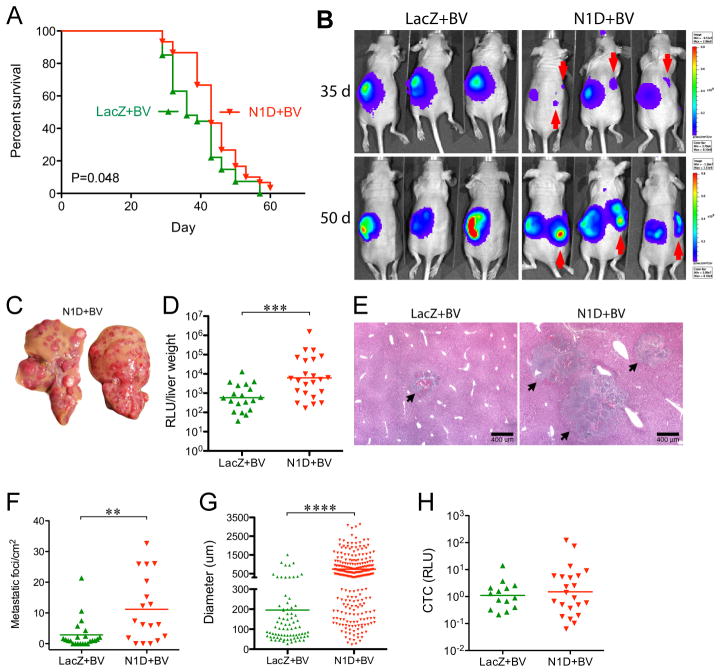
Figure 1. Combined Blockade of Notch and VEGF prolongs survival but increases liver metastasis.
(A) NGP-LacZ (n=30), or NGP-N1D (n=27) were treated with BV twice weekly, and sacrificed at a flux of 6x10⁹ photons/sec. P=0.048, Log-Rank (Mantel-Cox). (B) Bioluminescent images at days 35 and 50. Contralateral signal (red arrows) indicates metastatic disease. (C) Livers from NGP-N1D+BV mice display multiple nodules. (D) Liver metastatic burden was quantified by measuring bioluminescence of liver homogenates, NGP-N1D+BV (n=23), NGP-LacZ+BV (n=19). ***P<0.001. (E) Histologic examination of liver metastases (arrows). Bar, 400μm. (F) Metastatic foci/mm2, NGP-N1D+BV (n=18) NGP-LacZ+BV (n=23). **P<0.01. (G) Diameter of liver metastasis, with each point on the scatter plot an individual lesion. NGP-LacZ+BV (n=79, from 23 tumors); NGP-N1D+BV (n=254, from 18 tumors). ****P<0.0001. (H) Quantification of CTC. NGP-LacZ+BV (n=14), NGP-N1D+BV (n=22), P=n.s.

