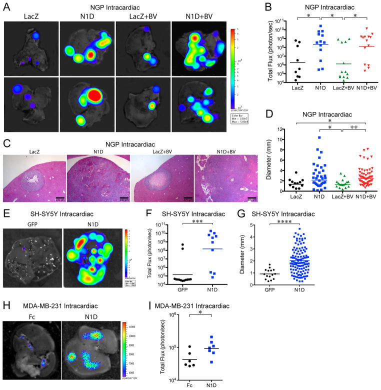
Figure 2. Blockade of Notch is sufficient to promote liver metastases.
NGP-LacZ or N1D cells were injected in the left ventricle, and treated with either placebo or BV; NGP-LacZ (n=9), NGP-N1D (n=13), NGP-LacZ+BV (n=13), NGP-N1D+BV (n=14). All mice were killed at 7 weeks. (A) Ex vivo imaging of livers. (B) Quantification of total flux (photon/sec) for NGP-LacZ (n=9), NGP-N1D (n=13), NGP-LacZ+BV (n=13), NGP-N1D+BV (n=14). *P<0.05. (C) Histologic examination of liver metastases. Bar, 400μm. (D) Quantification of liver metastases diameters: NGP-LacZ (n=12, from 9 tumors), NGP-N1D (n=35, from 13 tumors), NGP-LacZ+BV (n=17, from 13 tumors), NGP-N1D+BV (n=55, from 14 tumors). *P<0.05; **P<0.01. (E) Ex vivo imaging of livers, 6 weeks after intracardiac injection of SH-SY5Y-GFP (n=13) or SH-SY5Y-N1D (n=10). (F) Quantification of total flux (photon/sec) by ex vivo liver bioluminescence. ***P<0.001. (G) Quantification of liver metastases diameter for SH-SY5Y-GFP (n=15, from 1 tumor, 11 had none), SH-SY5Y-N1D (n=117, from 8 tumors, 3 had none). ****P<0.0001. (H) Ex vivo imaging of livers, 5 weeks after intracardiac injection of MDA-MB-231-Fc (n=8) or MDA-MB-231-N1D (n=9) (I) Quantification of total flux (photon/sec) by ex vivo liver bioluminescence. *P<0.05.

