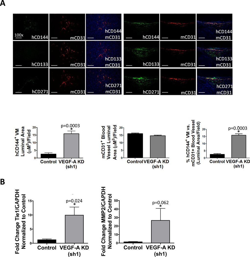
Figure 5. VEGF-A KD promotes VM and enriches MSLCs in WM1617 melanoma xenografts.
A, IF analysis of consecutive WM1617 xenograft sections double labeled for mCD31+ host blood vessels showed that WM1617 VEGF-A KD (sh1) xenografts displayed increased VM-engaging melanoma cells (hCD144+ in green; top panel) in close physical proximity to hCD133+ MSLCs (in green; middle panel) and hCD271+ MSLCs (in green; bottom panel) compared to nontarget control xenografts. Magnification, 100×. Scale bar: 200 µm. Human melanoma-derived VM vessels (hCD144+) and host angiogenesis (mCD31+) and were quantified as measured by the average luminal area per 100× field and revealed a significant increase in hCD144+ VM luminal area in WM1617 VEGF-A KD (sh1) xenografts compared to controls (left panel), no change in mCD31+ luminal area of host blood vessels (middle panel), and a significant increase in the percentage of melanoma VM channels (hCD144+) compared to mCD31+ blood vessels. *, P<0.05. C, Expression profiling by qRT-PCR revealed concomitant Tie-1 and MMP-2 upregulation in WM1617 VEGF-A KD (sh1) xenografts compared to nontarget controls. *, P<0.05.

