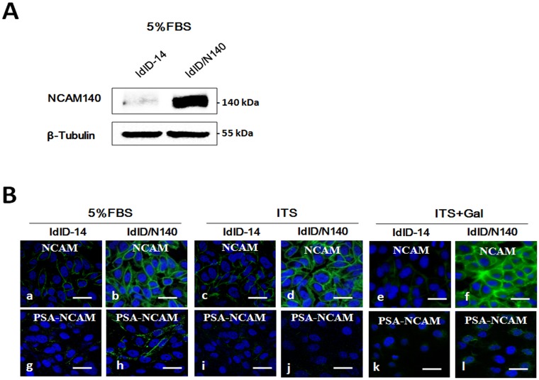
Fig 1. Overexpression of NCAM-140 in ldlD-14 cells.
(A) Western blot analysis with β-tubulin used as loading control. (B) Immunofluorescence staining of NCAM and PSA-NCAM. ldlD-14 and ldlD/N140 cells were cultured in serum-free medium supplemented with 5% FBS or ITS or ITS+Gal, and nuclei were visualized by Hoechst staining. Size bars: 20 μm.

