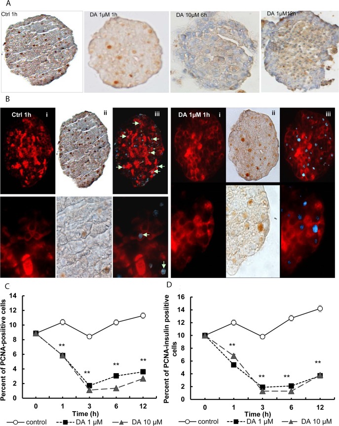
Fig 4. Effect of dopamine on cellular proliferation rate of insulin-positive beta cells in isolated islets.
(A) After double immunocytochemical staining, the nuclei of PCNA-positive cells appear brown (ii). (B) The proliferating insulin-positive cells (i, red) show brown-stained nuclei (ii), and the count of positive cells and calculation of the percentages of insulin-positive cells in proliferation was enabled after combining both images following digital transformation of the brown colour to blue colour (iii). 4000 cells per group were counted to obtain the statistical values. (C) Percentage of PCNA-positive cells out of the total number of cells (alpha, beta and delta cells), and (D) percentage of PCNA- and insulin-positive cells out of the total number of insulin-positive cells in the control pancreatic islets and those treated with 1μM and 10 μM dopamine **p<0.01.

