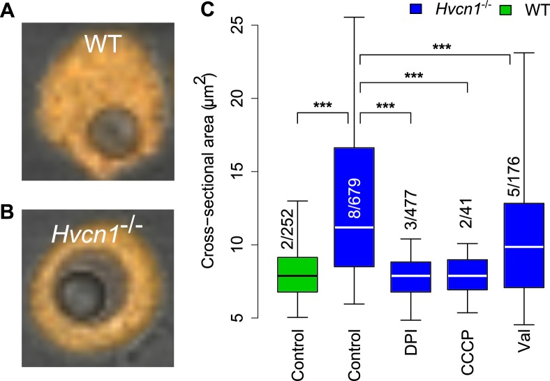
Fig 5. Vacuolar size in WT and Hvcn1 -/- neutrophils containing a single latex particle.
The cross-sectional area of a latex particle is ~7 μm2. Representative images of a WT (A) and Hvcn1 -/- neutrophil containing a single latex particle are shown in (A) and (B), respectively. (C) Quantitation of vacuolar swelling in Hvcn1 -/- neutrophils compared with WT, and the effects of 5 μM DPI, 60 μM CCCP and 3 μM valinomycin. The numbers of independent experiments is shown over the total number of measurements. Median, quartiles and 95% centiles are shown. Statistical significance: *** p < 0.001 and ** p<0.01.

