
Fig 8. The histological images of the cartilage of control and OA rabbits (HE staining, 20×).
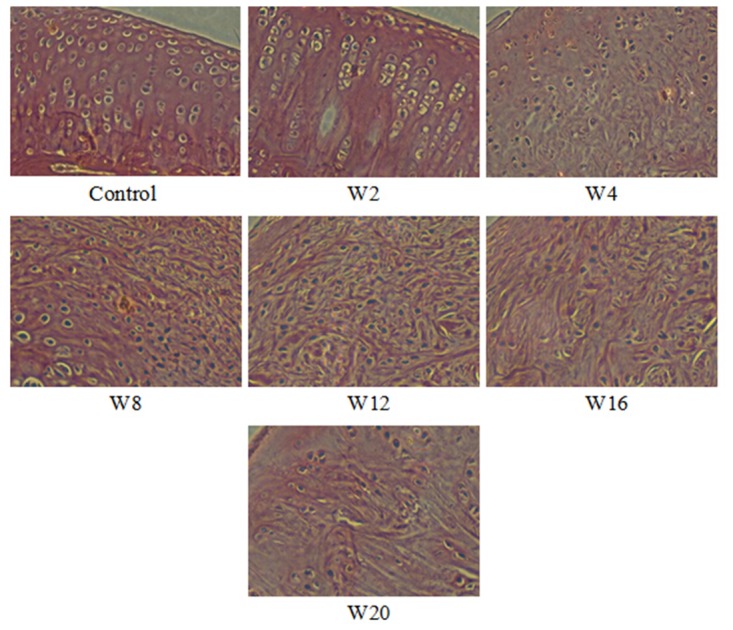
Control: Normal cartilage. W2–W20: The cartilage was subjected to the improved Hulth method and evaluated 2, 4, 8, 12, 16, and 20 weeks after surgery, respectively.