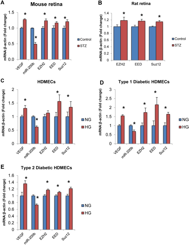
Fig 6. PRC2 components are altered in animal models of diabetes and other endothelial cell types exposed to high glucose.
A,B: Real time RT-PCR analysis of PRC2 component expression in animal retinal tissue from streptozotocin (STZ) induced diabetic and control animals. A: After 1 month of diabetes, VEGF expression was increased and miR-200b expression was decreased. Furthermore, EZH2, EED and SUZ12 levels were increased in rat retinal tissue from diabetic animals compared to control animals. B: After 2 months of diabetes, EZH2, EED and SUZ12 levels were increased in mouse retinal tissue of diabetic animals compared to controls. C,D,E: Real time RT-PCR analysis of VEGF, miR-200b and PRC2 components in HDMECs of various origins. In all groups, VEGF and miR-200b expression was altered. C: In non-diabetic adult HDMECs, EED and SUZ12 levels were significantly increased by HG. D,E: In HDMECs isolated from patients with diabetes (Type 1 & Type 2), EZH2, EED and SUZ12 were significantly increased in HG compared to NG controls. [HDMECs = human dermal microvascular endothelial cells, Type 1 = HDMECs isolated from a patient with type 1 diabetes, Type 2 = HDMECs isolated from a patient with type 2 diabetes, * p < 0.05 compared to control/NG; n = 6; data expressed as mean ± SEM, normalized to β-actin and expressed as a fold change of control].

