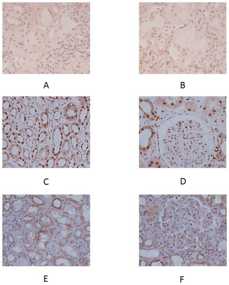
Fig 4. Expression of HMGB1 in renal tissue of active AAV patients and controls.

A and B showed isotype Ig staining in control renal tissue. Biopsy taken from normal renal tissue showed expression of HMGB1 mainly inside nuclei (C, D). Renal tissue of the active AAV patient showed strong cytoplasmic and extracellular staining for HMGB1 in tubuli and glomeruli (E, F).
