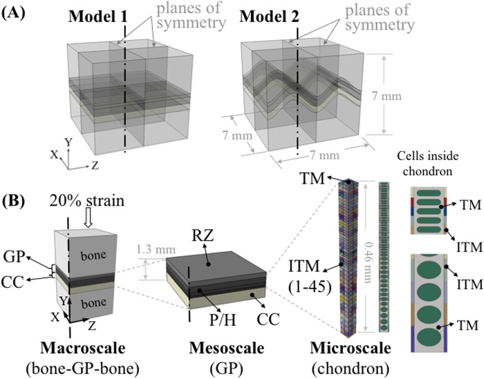
Fig 1. Overview of the multiscale modeling approach.
(A) Two idealized models were constructed consisting of epiphyseal bone, growth plate (GP) cartilage and metaphyseal bone (about 7 x 7 x 7 mm) with variations of mammillary processes: flat and ‘m’ shaped. (B) Set-up of the multiscale modeling approach. At the macroscale level, quarter models were used for analysis. About 0.69 mm thick growth plate cartilage was partitioned into two sections to represent the reserve zone (RZ) and the proliferative/hypertrophic (P/H) zone. Calcified cartilage (CC) was also included in the macroscale model. At the mesoscale, three individual layers were generated in the P/H zone to represent the gradient change of elastic modulus through the thickness of the growth plate. The microscale model of the chondron consisted of interterritorial matrix (ITM), territorial matrix (TM) and 46 chondrocytes with gradually changing cellular shape along with the same number of ITM sections. The elastic modulus of ITM increased from the RZ to the metaphyseal side to represent the gradual change of its material properties.

