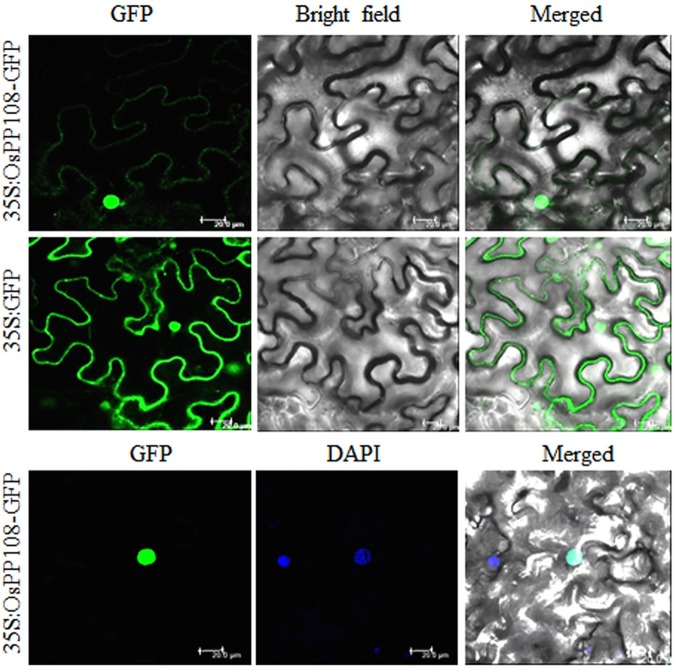
Fig 3. Sub-cellular localization of OsPP108 in Nicotiana benthamiana.
Agrobacterium-infiltrated tobacco leaves expressing the GFP-OsPP108 fusion protein driven by the 2XCaMV35S promoter. Confocal images of fluorescence (green) for cell expressing OsPP108 (35S:OsPP108-GFP) are showing its distribution predominantly in the nucleus and partly in cytoplasm (upper panel). Fluorescence is distributed throughout the cells, transformed with vector only (35S: GFP) (middle panel). Co-localization of green (GFP) signal with blue DAPI organelle marker confirms the localization of OsPP108 in nucleus (lower panel). Detection of fluorescence was performed under a confocal laser-scanning microscope (wavelength: 488nm for GFP and 405nm for DAPI). Scale bar = 20 μm.

