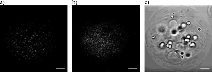
FIG. 6.
(a) Back-propagated SHG image of collagen fibers seeded at the core of a one day old MCF-7 core-shell Matrigel® + collagen + alginate bead. The image was acquired at a depth of 150 μm into the 300 μm diameter bead. (b) Maximum intensity z-projection of 31 optical sections acquired at 5 μm intervals from the top of the bead to 300 μm into the specimen. (c) A differential interference contrast image produced by collecting the forward propagated 810 nm excitation light after it passes through the specimen. In this image, the outline of the alginate shell, seeded cells and boundary of the Matrigel® + collagen + alginate core can be identified. Each scale bar represents 100 μm.

