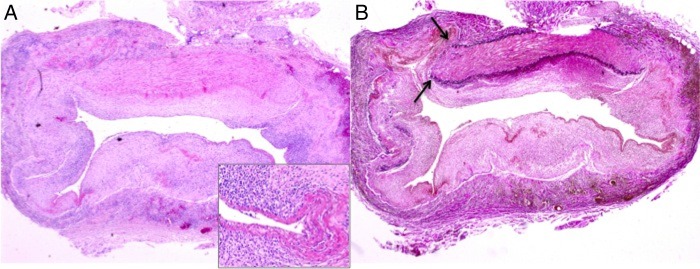
Figure 3.

Microscopic image of a medium-sized artery (H&E, original magnification ×2) demonstrating influx of inflammatory cells and fibrinoid necrosis of the arterial wall (inset) (A) with destruction of the internal and external elastic lamina (arrows; elastic van Gieson staining) (B).
