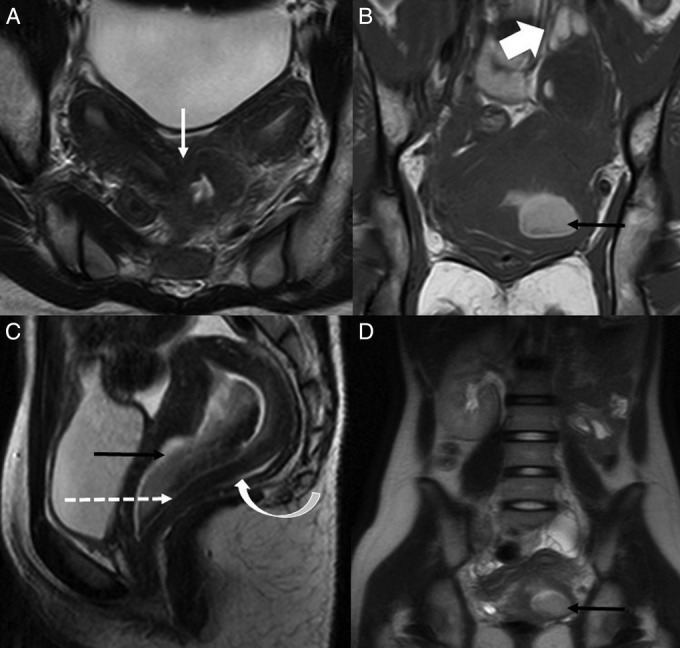
Figure 1.

(A) Axial T2-weighted MRI reveal two widely divergent, symmetrical uterine corpii having preserved zonal anatomy, partially fused at the level of cervix (white arrow) without any communication between their endometrial cavities. (B) Coronal T1-weighted and (C) Sagittal T2-weighted MRI shows a thick left hemivaginal septum (dashed arrow) separating the fluid filled dilated left hemivagina, which is hyperintense on T1-weighted and hypointense on T2-weighted images (black arrows), from the normally patent hemivagina of the right system (curved arrow). Note is made of a similar signal intensity tortuous dilated left haemosalpinx (broad white arrow) around the left ovary. (D) Coronal T2-weighted turbo spin echo image shows the true extent of the anomaly demonstrating the uterine didelphys with left haematocolpos (black arrow) and absent left kidney.
