Fig. 5.
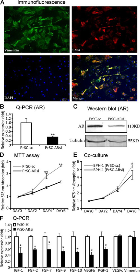
AR roles in the primary cultured prostate stromal cells. A: The immunofluorescence of vimentin (green) and SMA (red) in the primary cultured PrSCs. Scale bar = 100 µm. B: Reduced AR mRNA (left panel, Q-PCR) and (C) protein (right panel, Western blot) expression level in PrSC-ARsi cells, compared to PrSC cells. D: PrSC-ARsi cells showed lower growth rate than PrSC cells using MTT assay. E: BPH-1 cells co-cultured with PrSC-ARsi cells grew slower than those co-cultured with PrSC-sc cells using MTT assay. F: Decreased expressions of IGF-1, FGF-2, FGF-7, FGF-9, FGF-10,VEGFb, and PIGF, but not VEGFc and Vimentin, in PrSC cells with AR knockdown (**P < 0.01vs. PrSC-sc, *P < 0.05 vs. PrSC-sc; n = 3).

