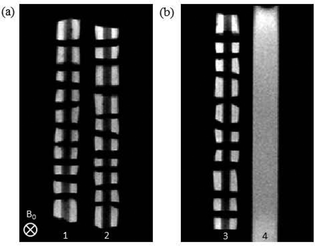
FIGURE 1.
Proton density weighted images (2D spin echo with TE = 10 ms) of the ULTEM sample holder containing ten BNC plugs per well (wells 1, 2, and 3), along with a DPBS standard (well 4) for intensity normalization of weighted images. BNC plugs were threaded onto hollow polyethylene tubes and separated by Teflon spacers. A single ROI consisted of two BNC regions, on either side of a polyethylene tube, from a single plug. These images indicate the slice orientation for each contrast modality. Note that the first slice bisected wells 1 and 2 (a), while the second bisected wells 3 and 4 (b).

