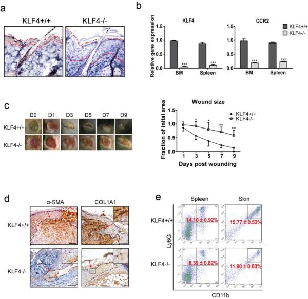
Figure 1. KLF4 ablation delayed cutaneous wound healing accompanied by decreased accumulation of MDSCs.
(a). KLF4 IHC staining in skin of RosaCreER/KLF4(flox) mice without (KLF4+/+) and with tamoxifen induction (KLF4−/−). The areas between two dotted red lines delineate skin suprabasal layers. (b). qRT-PCR to analyze expression of KLF4 and CCR2. (c). Wound healing phenotypes (left, n=10) and the wound size quantification (right). (d). IHC staining of α–SMA and COL1A1 in KLF4+/+ and KLF4−/− mice at Day 3 after wound induction. (e). Flow cytometry using anti-Ly6G and anti-CD11b antibodies with spleen cells and single skin cells. Representative images from one of five mice in each group are shown (a, c, and d). Scale bars: 50 μm. Mean ± SEM. *p< 0.05, **p< 0.01, ***p< 0.001.

