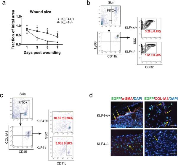
Figure 2. Delayed wound healing in bone marrow KLF4 knockout mice and compromised accumulation of CCR2+MDSCs and fibrocytes.
(a). Chimeric mice receiving bone marrow cells from Rosa26CreER/KLF4(flox)/β-actin-EGFP donor mice were used. Quantification of the wound size in each group of mice is shown (n=10). (b). Single cells from the skin wound were gated by EGFP. They were examined by CD11b and Ly6G antibodies, followed by further analysis using a CCR2 antibody. Representative contour plots in each group are shown. (c). Similar to (b) except COL1A1, CD45, and CD11b antibodies were used to analyze the fibrocytes. (d). Representative immunofluorescent staining of the wounds with α-SMA and COL1A1 antibodies. Yellow arrows indicate EGFP/α-SMA or EGFP/COL1A1 co-expressing cells (Scale bars: 50 μm).

