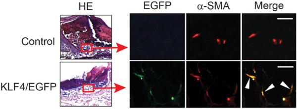
Figure 3. KLF4-expressing bone marrow cells are integrated into the healing tissue and co-localized with α-SMA-expressing cells.

Chimeric mice were generated by bone marrow transplantation using bone marrow cells from C57BL/6 mice (Control) or KLF4/EGFP mice into C57BL/6 recipient mice. 8mm diameter full thickness wound was placed and the wound beds were collected 4 days later followed by immunofluorescence staining with an anti-α-SMA antibody. Representative co-localization of EGFP cells with red α-SMA-expressing cells in the healing tissue (left) are indicated by white arrowheads (n=5). Scale bars: 25 μm.
