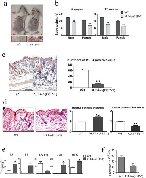
Figure 4. Hair loss and decreased fibrocyte generation in FSP-1-Cre/KLF4(flox) mice.
(a). Representatives of the wild type (WT) and FSP=1-Cre/KLF4(flox) (KLF4−/−(FSP-1)) mice. Black squares indicate an area in which a severe hair loss was seen in KLF4−/−(FSP-1) mice. (b). Body weights of male and female WT and KLF4−/−(FSP-1) mice. (c). Representative images of KLF4 staining of skin (left) and measurement of KLF4 positive cells (right). (d). Left, representative images of HE staining of skin. The areas between two dotted red lines represent skin suprabasal layers and the red arrow heads are pointing to hair follicles. Right, measurement of epithelial thickness and hair follicles. (e). Measurement of serum cytokines by ELISA (n=3). (f). Quantification of fibrocyte generation (n=5) Scale bars: 100 μm. *p<0.05, **p<0.01.

