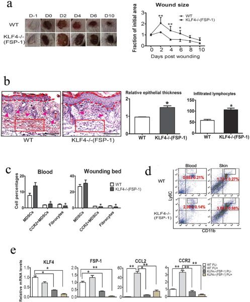
Figure 5. Compromised wound healing of PU in FSP-1-Cre/KLF4(flox) mice associated with decreased CCR2+MDSCs and fibrocytes.
(a). Similar to Figure 1c, except the WT and KLF4−/−(FSP-1) mice were used in the PU model (n=10). (b). Left: Representative images of HE staining of skin wounds in PU model. Right: Quantification of the epithelial thickness and the numbers of infiltrated lymphocytes in arbitrary red squares with the same sizes as the left (n=5). (c). Flow cytometry analysis to examine MDSCs and fibrocytes in mouse blood and skin wounds. (d). Similar to c, CD11b and Ly6C antibodies were used to examine inflammatory monocytes. (e). qRT=PCR to analyze expression of KLF4, FSP-1, CCL2, and CCR2 in the skin wounds. Scale bars: 100 μm, *p<0.05, **p<0.01.

