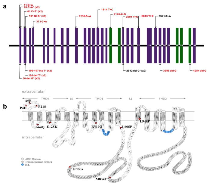
Figure 1. The positions of ABCC6 mutations identified in Chinese patients with PXE.
(a) Intron-exon organization of ABCC6 gene. Vertical boxes represent the 31 exons; Missense mutations are shown above, and insertion or deletion mutations resulting in PTC below the line; Green exons code for the two nucleotide-binding fold domains of the protein; Black, previously reported mutations; Red, to our knowledge previously unreported mutations; *denotes the presence of the mutation in multiple alleles/patients with the number of affected alleles in parenthesis. (b) Positions of the missense variants in the membrane topology model of the ABCC6 protein. The various protein domains are delineated by horizontal arrows above; the positions of amino acid variants investigated in the study are in red; nucleotide binding fold domains and intracellular loops are colored with gray and blue, respectively.

