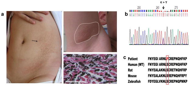
Figure 4. Cutaneous presentation, histopathology and mutation detection in a pediatric patient with PXE.

(a) Hyperpigmentation on the trunk (left) and yellowish papules in the axillary fossa (upper right); Aberrant calcification in the dermis detected by von Kossa stain (bottom right); (b) A heterozygous mutation, p.S479F, in the ENPP1 gene revealed by mutation analysis (arrow); (c) Conservation of the serine-479 during evolution from zebrafish to human (outlined). Scale bar = 100 mm.
