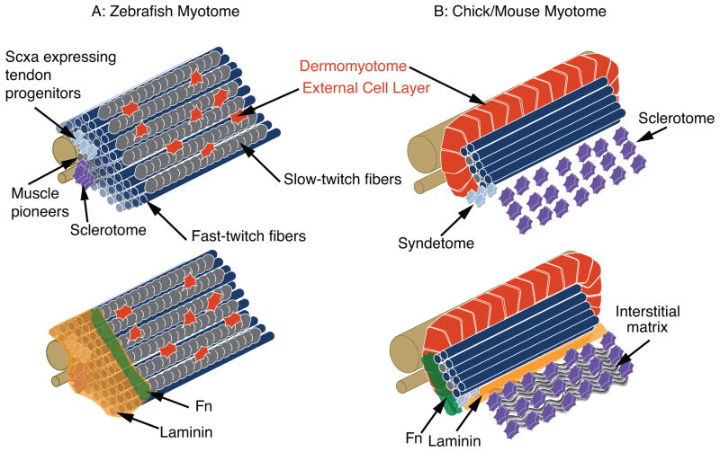
Figure 2.
Structure of the zebrafish and amniote myotomes. A: Top Panel - Muscle is the major constituent of the zebrafish myotome. Tendon progenitors and sclerotome are located medially. Most of the muscle cells are fast-twitch muscle. The most superficial muscle fibers are slow-twitch muscle fibers (gray). The external cell layer (red) is hypothesized to be somewhat equivalent to the amniote dermomyotome. Bottom panel - The ECM at the MTJ is superimposed upon a myotome. Laminin is expressed throughout the medial-lateral extent of the MTJ, but Fn is degraded medially to migrating slow-twitch fibers to end up primarily concentrated at the MTJ adjacent to slow-twitch fibers. B: Top Panel - Structure of the amniote myotome. The epithelial dermomyotome contains muscle progenitor cells that will sustain growth and will also give rise to satellite cells. The connective tissue progenitor region is termed the syndetome. Bottom panel - ECM of the amniote myotome. Note that the myotomal BM separates the sclerotome from the myotome. Fn is primarily concentrated at myotome boundaries.

