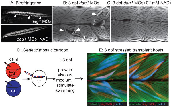
Figure 5.

An organized ECM microenvironment rescues fiber resiliency in Dag1-deficient cells (from Goody et al., 2012). Anterior left, dorsal top, side-mounted, 3 dpf embryos. A: Polarized light microscopy shows loss of birefringence in dag1 morphant myotomes (white arrowheads). Birefringence is rescued in NAD+-supplemented dag1 morphants. B: 3 dpf embryos stained with phalloidin (white). Fiber detachment is readily observed in dag1 morphants (arrowheads). C: NAD+ supplementation reduces fiber detachment in dag1 morphants. D: Genetic mosaic cartoon depicting transplantation of fluorescent dextran-labeled dag1 morphant (red) and control (blue) cells into unlabeled, control hosts. Embryos were stressed (frequently stimulated to swim in a viscous medium), and reared to 3 dpf. E: Transplanted control cells (blue) and dag1 morphant cells (red) remain attached to MTJs, even when hosts are stressed. This suggests that a normal host ECM microenvironment is sufficient for resiliency of dag1 morphant cells and supports that NAD+ functions via augmentation of the ECM microenvironment.
