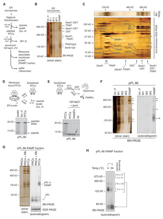
Figure 1. Nascent pPL-86 Binds to a Subset of ER-Derived RAMPs.
(A) Schematic of protocol used to isolate ribosome-associated membrane proteins (RAMPs). (B) Blue native (BN) - PAGE analysis of RAMPs labeled with translocon complexes. (C) 2nd dimension SDS-PAGE analysis of a BN-PAGE gel slice (silver stain) containing translocon components and RAMP complexes as labeled. Asterisks denote TRAP components in the 750 kD RAMP complex. (D) pPL-86 ribosome-translocon complexes (RTCs) were solubilized and pelleted (pel) by ultracentrifugation and analyzed by SDS-PAGE (autoradiogram). Cleared lysate (lys, cl) shows little pPL-86 remaining in the supernatant (sup). (E) SDS-PAGE (autoradiogram) of pPL-86 RTCs before and after NaCl and puromycin (puro) treatment followed by fractionation using ultracentrifugation into RAMP fractions (arrow) and ribosome pellets (rib). (F) BN-PAGE of cleared lysate, wash (W), and RAMP fractions obtained from pPL-86 in vitro translation reactions. (G) pPL-86 ribosome-nascent chain complexes with or without addition of solubilized membranes (RNCs or RNCs + Mb) did not associate with RAMP complexes on BN-PAGE. (H) Incubation of pPL-86 RAMP complexes at 24°C prior to BN-PAGE. Percent nascent chain remaining associated with each RAMP complex after incubation is shown (± SEM, n=3). See also Figure S1.

