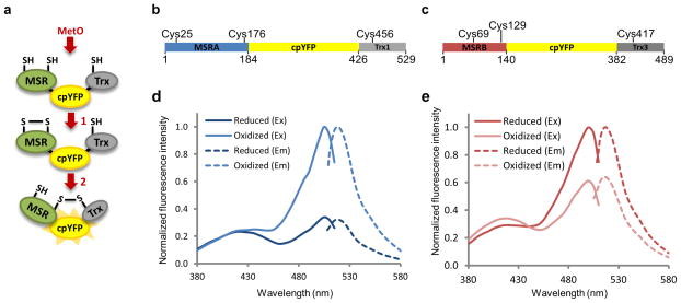
Figure 1. Design and spectra of MetSOx and MetROx sensors.
(a) Mechanism-based design of MetO sensors. 1) After reaction with MetO, disulfide is formed between two redox-active Cys of an MSR moiety. 2) Reduction of this bond by the catalytic Cys of the Trx moiety induces a conformational change in the sensor leading to changes in spectral properties of cpYFP. (b) Schematic representation of MetSOx, the sensor specific for the S-diastereomer of MetO. cpYFP (yellow) was fused in frame with yeast MSRA (blue) and yeast Trx1 (grey). C25, C176 and C456 represent redox-active Cys residues involved in catalysis. (c) Schematic representation of MetROx, the sensor specific for the R-diastereomer of MetO. cpYFP (yellow) was fused in frame with yeast MSRB (red) and yeast Trx3 (dark grey). For both MSRB and Trx3, mitochondrial targeting sequences were removed. C69, C129 and C417 represent redox-active Cys residues involved in catalysis. Excitation (Ex) (full line) and emission (Em) (dashed line) spectra of reduced (dark color) and oxidized (light color) MetSOx (d) and MetROx (e). Spectra were normalized to the oxidized form. The data presented are representative of 3 replicates. Sensors were reduced with 10 mM DTT and oxidized with 10 μM MRP4.

