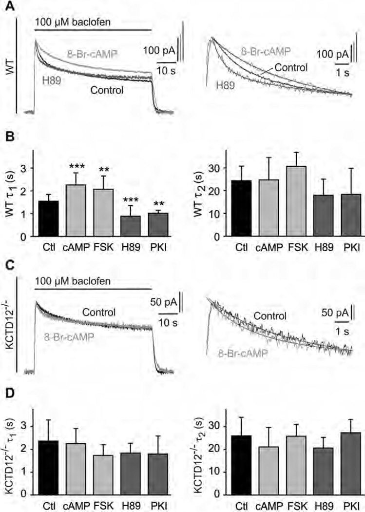
Fig. 4.
PKA activation slows fast desensitization of baclofen-activated K+ currents in cultured hippocampal neurons of WT but not of KCTD12−/− mice. (A) Representative baclofen-activated K+ currents recorded at −50 mV from WT hippocampal neurons. PKA activity was modulated by pre-incubation for 30 min with 8-Br-cAMP (1 mM; bright grey trace) or H89 (2 µM; dark grey trace). Controls represent recordings from untreated neurons (black trace). The desensitization time constants τ1 and τ2 were derived from double-exponential fits to the decay phase of K+ currents during baclofen application (enlarged on the right). (B) Bar graph summarizing the time constants τ1 and τ2 of baclofen-induced K+ current desensitization for the indicated treatments. Data are means ± SD, n = 7–35. **, p < 0.01; ***, p < 0.001; Dunnett’s multiple comparison test, compared to Ctl. Ctl, control; FSK, forskolin; cAMP, 8-Br-cAMP. (C) Representative baclofen-activated K+ current responses from hippocampal KCTD12−/− neurons pre-incubated with 8-Br-cAMP (grey trace) or untreated KCTD12−/− neurons (control, black trace) as in (A). Traces were fitted as in (A). (D) Bar graph summarizing the time constants τ1 and τ2 of baclofen-induced K+ current desensitization for the indicated treatments. Data are means ± SD, n = 5–17, Dunnett’s multiple comparison test, compared to Ctl.

