Figure 1.
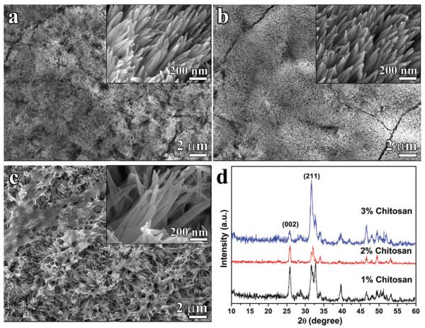
(a–c) SEM images of the newly grown layer after remineralization in CS-AMEL hydrogel with 1% m/v (a), 2% m/v (b) and 3% m/v (c) chitosan. Insets show the crystal morphology at high magnification. (d) XRD patterns of the newly grown layer after remineralization in CS-AMEL hydrogel with different concentrations of chitosan.

