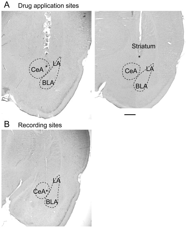
Figure 10. Histologic images of drug application and recording sites.
A, Individual examples of histologically verified positions of microdialysis probes in the central nucleus of the amygdala (left) and striatum (placement control, right). Asterisks denote position of the tip of the microdialysis fiber which protruded the shaft of the probe by 1 mm. Note that the shaft left a larger lesion than the fiber. Removal of the probe and processing of the tissue likely increased the size of the lesion. B, Histologically verified position of the recording electrode in the capsular division of the central nucleus. Asterisk denotes position of the electrode tip. CeA, central nucleus of the amygdala; LA, BLA, lateral and basolateral amygdala. Calibration bar, 1 mm.

