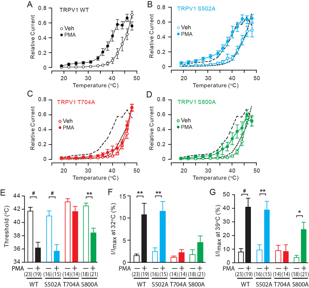
Fig. 1. The roles of phosphorylation sites in PKC-induced hypersensitivity of TRPV1 to heat in HEK293 cells.

A–D. Averaged relative current density evoked by a temperature ramp from 17 to 48 °C following the exposure to either vehicle (white) or 1 µM PMA (black) in HEK 293 cells transfected by TRPV1 WT (A), S502A (B), T704A (C), or S800A (D). Current amplitudes measured at −50 mV at different temperatures were normalized to the maximal amplitude in each cell. Black dotted lines in B–D represent values of WT.
E–G. Quantification of heat-evoked currents in TRPV1 WT and mutants. Threshold temperature of activation (E), relative currents at 32°C (F) and 39°C (G) were compared between vehicle-treated (open bars) and PMA-treated (filled bars) cells as indicated. Relative currents at 31°C or 39°C were calculated by averaging current amplitudes measured between 29°C to 34°C or 38°C to 40°C in each cell and normalizing to the respective maximal current amplitude in the given cell. Numbers within parenthesis represent the numbers of observations. *p<0.05; **p<10−3; #p<10−4; Student’s t-test.
