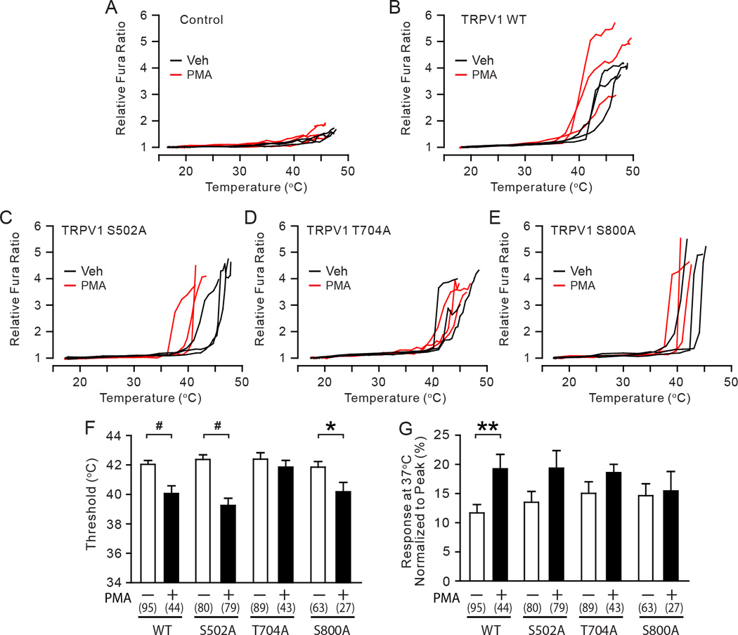
Fig. 5. The roles of phosphorylation sites in PKC-induced hypersensitivity of TRPV1 to heat in sensory neurons.

A–E. Representative traces of the relative Fura responses evoked by temperature ramps from 16 to 48 °C following the exposure to either vehicle (black) or 0.3 µM PMA (red) in TRPV1 null DRG neurons transfected by an empty plasmid (A), TRPV1 WT (B), S502A (C), T704A (D), or S800A (E). The Fura ratio was normalized to the minimum value measured at cold temperature range in each cell.
F–G. Quantification of heat-evoked responses in TRPV1 WT and mutants. Threshold temperature of activation (F) and the relative response at 37°C (G) were compared between vehicle-treated (white bars) and PMA-treated (black bars) cells as indicated. The relative responses at 37°C were calculated by averaging the relative Fura responses measured between 36°C to 38°C in each cell and normalizing to the respective peak response in the given cell. Numbers within parenthesis represent the numbers of observations. *p<0.05; **p<0.01; #p<0.0005; **p<0.01; Student’s t-test.
