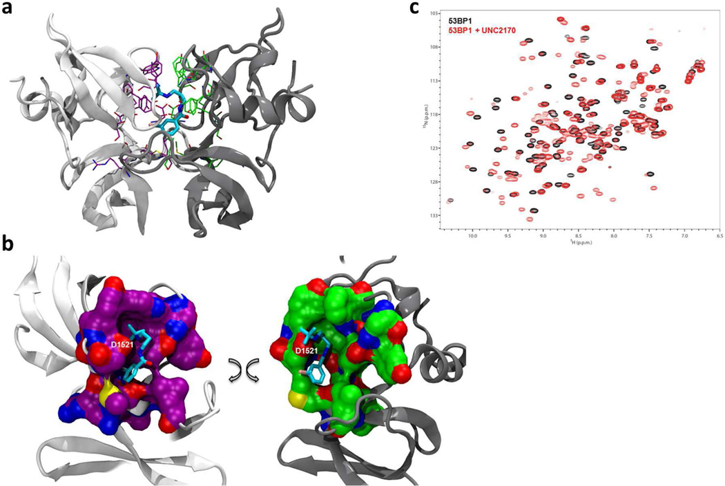
Figure 2.
Crystallography and NMR evidence for UNC2170 (1) binding to 53BP1. (a) Co-crystal structure of 1 (cyan) bound to the 53BP1 tudor domain dimer (PDB 4RG2). One 53BP1 protein unit is shown in light gray and the other in dark gray, with the residues that interact with UNC2170 shown in fuchsia and green, respectively. (b) View of the protein surface of each 53BP1 tudor domain that interacts with UNC2170 (the two domains shown in (a) are separated and rotated); color coding is the same as in (a). (c) Overlay of the 1H-15N HSQC correlation spectra of 53BP1 in the free state (black) and in the presence of 10-fold molar excess UNC2170 (red).

