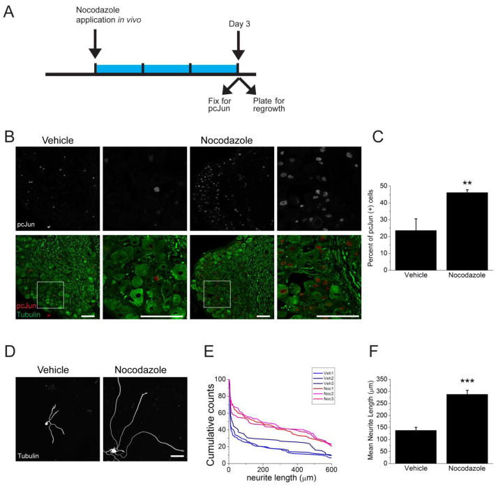
Figure 8. Nocodazole application in vivo induces pcJun upregulation and enhances axonal regrowth.
A. Schematic diagram outlining the in vivo experiment. Nocodazole was applied to the sciatic nerve for 3 days (blue bar) prior to dissection of DRGs for pcJun and regrowth assessment.
B. Sample confocal images of sections of adult DRGs stained for pcJun (red) and β3 tubulin (green). Application of nocodazole to the sciatic nerve significantly increases the number of neurons with pcJun positive nuclei. Boxed areas are shown at higher magnification in the adjacent panels. Scale bar = 100 μm.
C. Percentage of pcJun positive cells in the groups represented in B. **p<0.01, n = 3 mice.
D. In vivo application of nocodazole to the sciatic nerve prior to dissociating and plating neurons enhances the regrowth compared to vehicle treatment. Sample confocal images of dissociated DRG neurons 18 hours after plating stained for β3 tubulin. Scale bar = 100 μm.
E. Representative cumulative distribution plots for the longest neurite per cell from each mouse. Red lines represent mice treated with nocodazole and blue lines represent vehicle-treated animals. ~100 neurons were measured for each mouse.
F. Mean neurite length was measurement for each mouse represented in E. ***p<0.001.

