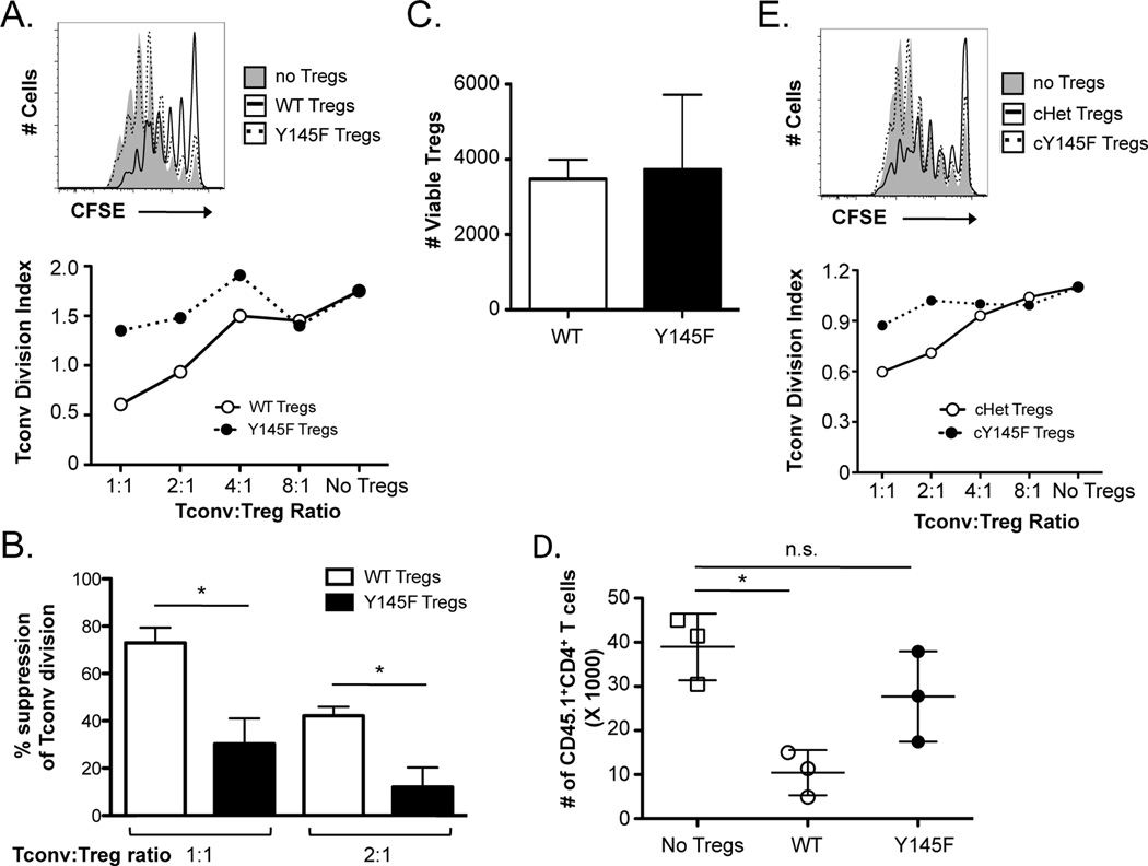
Figure 2. SLP-76 Y145F Tregs demonstrate greatly diminished in vitro and in vivo suppressive function.
(A) CD4+CD25+ Tregs were sorted from SLP-76 Y145F mice and co-cultured with CFSE-labeled CD4+Foxp3− cells (Tconvs) at various ratios for four days in the presence of irradiated T cell-depleted splenocytes and anti-CD3. A representative CFSE plot from cultures lacking Tregs or at a 1:1 Treg:Tconv ratio is shown (top). A representative plot of the division index of Tconvs at various Treg:Tconv ratios is shown (bottom). (B) The percent suppression at a 1:1 and 1:2 Treg:Tconv ratio were compiled from 3 independent experiments and represented as mean ± SEM. (C) The absolute number of live WT and SLP-76 Y145F Tregs from 3 independent experiments was determined using a viability dye in the 4-day co-cultures. (D) TCRβ/δ DKO mice were adoptively transferred with Tconvs with or without Tregs from B6 or SLP-76 Y145F mice. The absolute number of Tconvs recovered is shown. One representative of two independent experiments is shown. (E) YFP+CD4+CD25+ Tregs from Tamoxifen-treated SLP-76 cHet or SLP-76 cY145F mice were co-cultured with CFSE-labeled Tconvs for four days in the presence of irradiated T cell-depleted splenocytes and anti-CD3. A CFSE plot from cultures lacking Tregs or at a 1:1 Treg:Tconv ratio (top) and a plot of the division index of Tconvs at various Treg:Tconv ratios are shown (bottom). One representative of 2 independent experiments is shown. * indicates statistical significance of p < 0.05 by unpaired two-tailed Student t-test.

