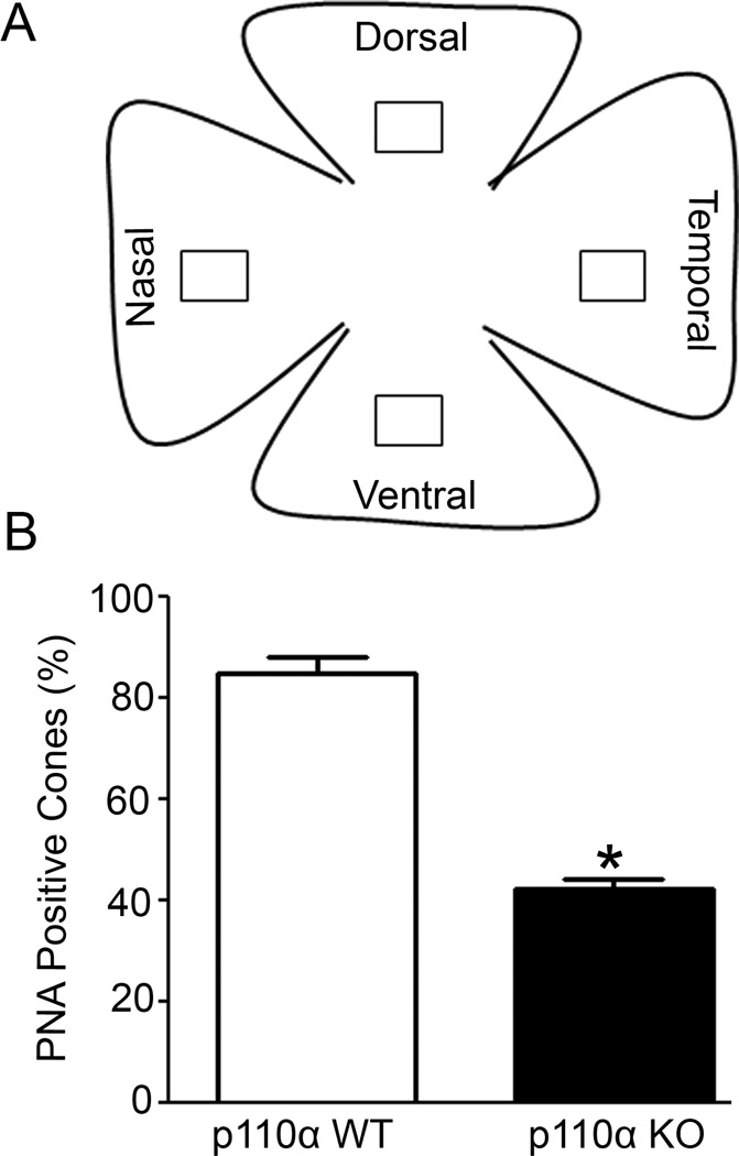
Figure 4. Quantification of cone cell loss in cone photoreceptors in p110α KO retinas.
PNA-labelled retinal whole mounts from WT and p110α KO mice were subjected to imageJ analysis on selected areas (□) of dorsal, ventral, nasal and temporal regions of the retina (A). Combined PNA-labeled positive cones were expressed as percentage and the wild-type was set as 100 percent (B). Data are mean ± SEM, n = 3, *p<0.001.

