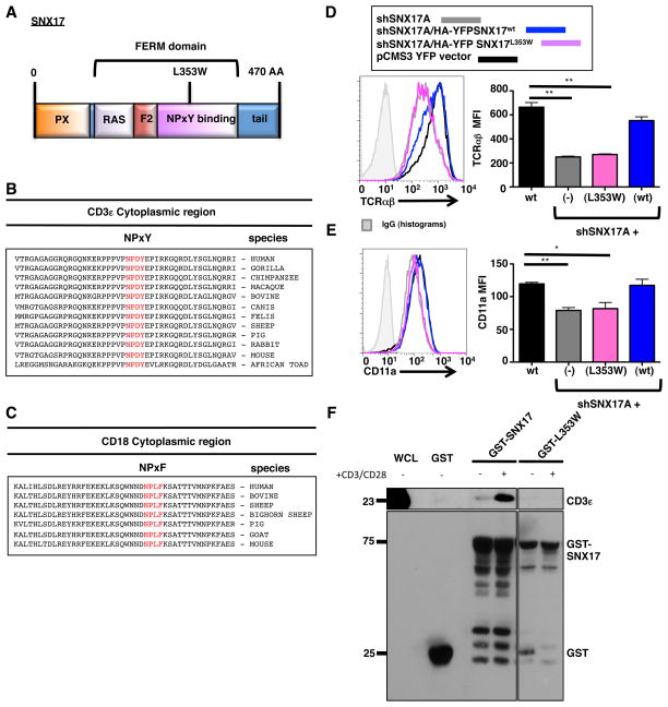
Figure 7. The SNX17 FERM-domain binds and traffics TCR and LFA-1.
(A) Diagram of SNX17. (B) Peptide alignment of the cytoplasmic region of CD3ε. (C) Peptide alignment of the cytoplasmic region of CD18. (D) Transfected pCMS3. YFP vector control (black), shSNX17A (gray), shSNX17A/HA-YFP SNX17WT (blue), and shSNX17A/HA-YFP SNX17L353W (magenta) Jurkat T cells were surfaced labeled for TCRαβ and YFP+ cells were analyzed using flow cytometry. (E) Transfected pCMS3 YFP vector control (black), shSNX17A (gray), shSNX17A/HA-YFP SNX17wt (blue), and shSNX17A/HA-YFP SNX17L353W (magenta) Jurkat T cells were surfaced labeled for CD11a and YFP+ cells were analyzed using flow cytometry. (F) GST-pull down assay using whole cell lysates from unstimulated or anti-CD3/CD28 treated primary human CD4+ T cells. The pull-down was performed with GST only control, GST-SNX17, or GST-SNX17 (L353W) mutant and immunoblotted for CD3ε and GST. The results in D and E are representative of four separate experiments, while F is representative of 3 separate immunoblot experiments. Bars represent mean ± SEM. Horizontal lines indicate statistical comparison between groups, *p≤0.05, **p≤0.005.

