
Figure 3.
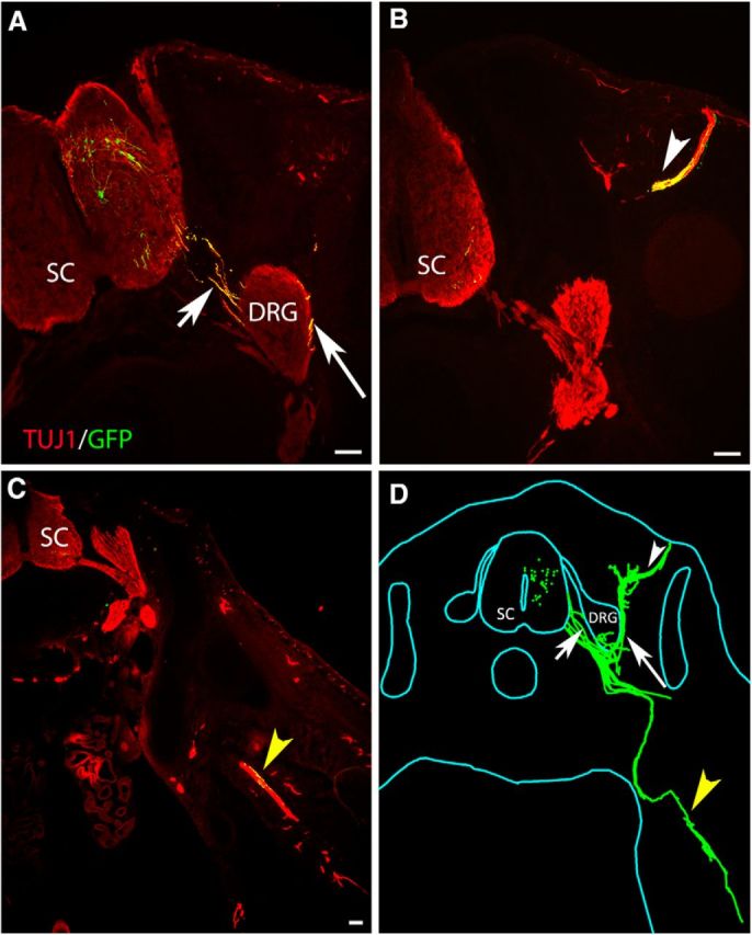
iPSCMNs preferentially project axons to epaxial muscles when transplanted in ovo. A–C, Cross-section through a HH St. 31 chick embryo showing eGFP+ motor axons extended out of the spinal cord through the ventral root (A, short arrow). The majority of the eGFP+ axons extended around the DRG (A, arrow) and into epaxial muscles (B, white arrowhead). A few eGFP+ axons extended ventrally into the limb bud (C, yellow arrowhead). All sections were immunolabeled with Tuj1 to visualize the endogenous chick neurons (red) and transplanted eGFP+ iPSCMNs (yellow). D, Neurolucida reconstruction of all cross-sections from one chick embryo receiving an iPSCMN transplant. eGFP+ iPSCMNs are shown in green. Scale bar, 100 μm.