
Figure 1.
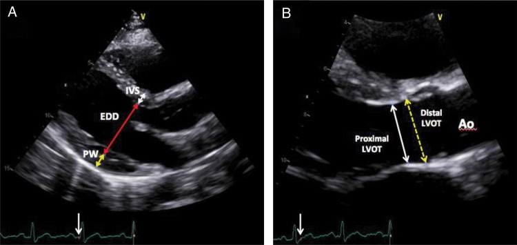
(A) Two-dimensional-guided measurement of left ventricle wall thickness in end-diastole from the left parasternal long-axis view. The interventricular septum thickness (white arrow), the left ventricle end-diastolic diameter (red arrow) and the posterior wall (PW; yellow arrow) thickness are measured just distal to the mitral leaflets tips, perpendicular to the long axis of the LV. (B) Proximal left ventricle outflow tract (LVOT) diameter was measured in mid-systole, using the trailing-edge-to-leading-edge method, 0.5–1 cm below the aortic cusps in a plane parallel to the aortic annulus (white arrow) from the zoomed parasternal long-axis view. The yellow dashed arrow represents the distal LVOT diameter measured just below the aortic annulus level.