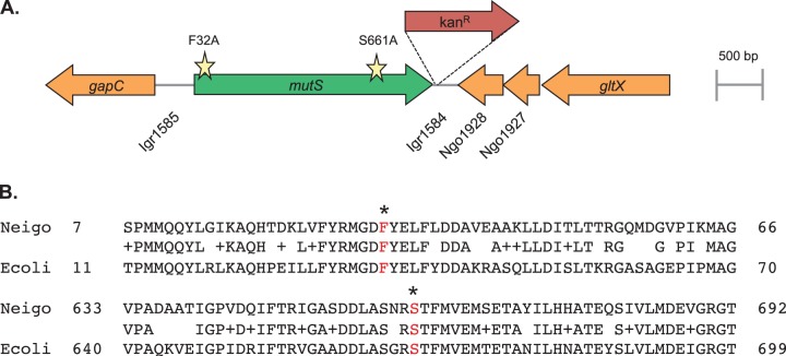
FIG 2.
The mutS locus of N. gonorrhoeae. (A) Schematic representation of the mutS locus in N. gonorrhoeae strain FA1090 with the Kanr marker used to introduce the site-directed mutations. Open reading frames are indicated by arrows drawn in the direction of transcription. Gene or locus names are written either inside the arrow or directly underneath. The lines between genes represent intergenic regions (Igr). Stars represent the locations of the F32A and S661A amino acid substitutions. Drawings are to scale. (B) Alignment of N. gonorrhoeae (Neigo) MutS and E. coli (Ecoli) MutS amino acid sequences. Identical and related residues are shown in the center line, and the conserved phenylalanine and serine residues (in red) are indicated with an asterisk.

