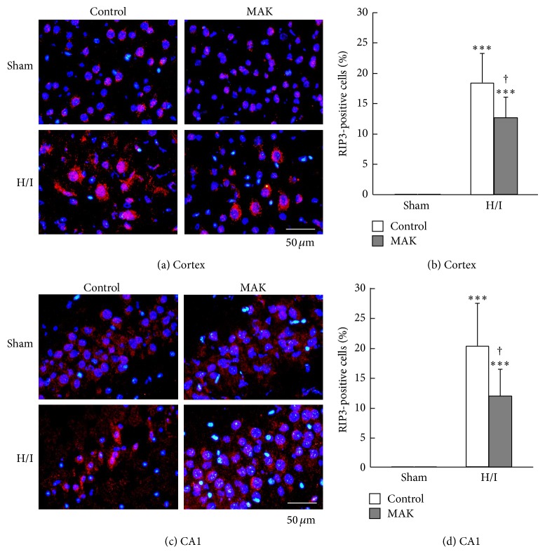
Figure 7.
Effects of chronic pretreatment with MAK (1 g/kg) on RIP3 expression in the ischemic penumbral regions. (a) Representative photographs of RIP3 immunostaining at 24 h of reoxygenation after H/I in the penumbral cortex (a) and hippocampal CA1 region (c) from the mice in each group. Scale bar = 50 μm. Quantification of the number of RIP3-positive cells was achieved by cell counting in the penumbral cortex (b) and hippocampal CA1 region (d) from the mice in each group. Throughout, data are represented as means ± S.D. from 3–5 mice in each group. ∗∗∗ P < 0.001 compared with the respective sham-operated groups. † P < 0.05 compared with the H/I-treated control group.

