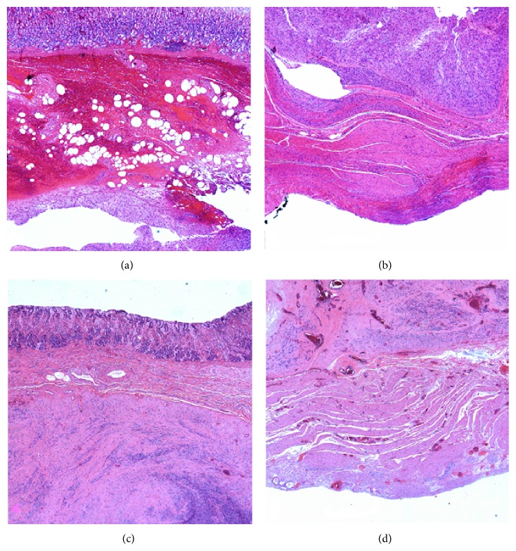
Figure 3.
Patient 3's endoscopic specimen (a) shows spindle cells representative of GIST involving the submucosa and margin of the sample. The laparoscopic specimen (b) from the same patient demonstrates GIST cells confined superficial to the serosal surface. Patient 4's endoscopic specimen (c) likewise shows spindle cell involvement at the specimen's margin, while the laparoscopic specimen (d) exhibits a negative oncologic margin.

