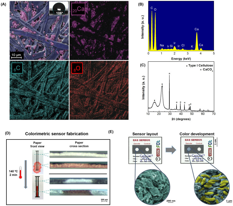
Figure 3. Office paper characterization.
(A) SEM image and EDS map; (B) EDS spectrum; (C) XRD diffractogram; (D) Hydrophobic barriers formation; (E) Photograph of a positive result in the developed paper-based sensor with WO3 nanoprobes for the colorimetric detection of EAB (Geobacter sulfurreducens cells in yellow and hexagonal WO3 nanoparticles in blue). The images are false-colored (GIMP software) for better understanding of the different materials.

