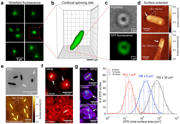
Figure 1. Single cell surface adhesion and irreversible attachment on continuously growing S-EPS areas.
(a) Ex-vivo WFM images of individual surface-adhered Xylella fastidiosa by their polar region. (b) Ex-vivo SDCLM images of reversibly adhered bacteria via the polar region. (c) Brightfield and corresponding WFM images reveal circular S-EPS structures at bacterial adhesion regions. (d) SPM images show differences in surface potential (Δ = ~100 mV) indicating S-EPS coating at the polar regions of the bacteria. (e) Contrast difference in SEM image and changes in surface potential (Δ = ~70 mV) identify S-EPS disks around irreversibly attached bacteria. (f) Fluorescence staining (PAS, periodic-acid-Schiff) of polysaccharides show growing circular S-EPS shapes over time. (g) Histogram of S-EPS disks covered area (n = 168 for each growth time) demonstrates disk growth with increasing bacteria incubation time (example false-color fluorescence images on left panel for the different growth times of 6, 12 and 18 h). See also Supplementary Figs. S1 and S2. Measurement statistics are described in the Materials and Methods section.

