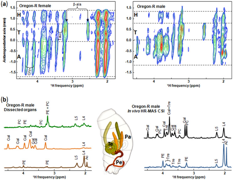
Figure 2. HR-MAS CSI experiments.
(a) In vivo1H 2D HR-MAS CSI spectra of female (left, body length of ~ 3 mm) and male (right, body length of ~ 2.5 mm) Oregon-R drosophila. In the 2D maps, the regions corresponding to the head (H), thorax (T) and abdomen (A) are separated by the dashed lines. Sixteen contour levels are plotted with a top contour of 5% of the maximum intensity ((CH2)n resonance at 1.3 ppm) and a dividing factor of 1.22. β-ala: β-alanine; Tau: Taurine; Gly: glycerol. (b) Left:1H 1D HR-MAS spectra of dissected organs of 2 to 5 Oregon-R males. Green, orange and brown spectra correspond to testis, paragonia and penis, respectively. Middle: Scheme of the male reproductive system (dorso-lateral view oriented anterior top, from reference 24). Te: testis, Pa: paragonia, Pe: penis apparatus. Right: 1D sum along the spatial dimension (vertical) of the 2D 1H HR-MAS CSI spectrum of the male fly for the two specific regions corresponding to the middle (black) and posterior (blue) parts of the abdomen. Ac, acetate (or acetyl); Gal, galactoside; L4, CH2C = ; L5, CH2CO; PC, phosphocholine; PE, phosphoethanolamine; Tre, trehalose. All spectra were recorded at a magnetic field of 17.6 T. The spinning frequency was 2630 Hz for in vivo experiments and 4000 Hz for dissected organs.

