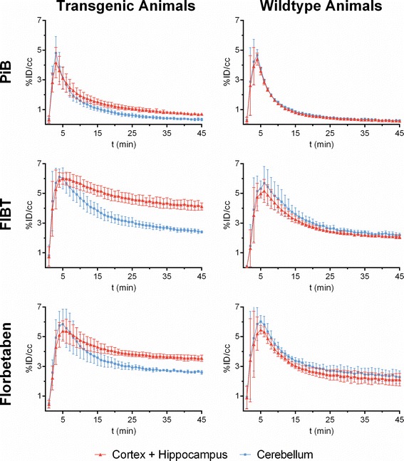
Figure 3.

Dynamic PET time activity curves of the cortex-VOI and the cerebellum-VOI. Time-activity curves of the three radiopharmaceuticals (rows) within four APP/PS1 tg mice (left column) and three control mice (right column). Values are percentage of injected dose per cubic centimeter (%ID/cc) ± SD for a cortex + hippocampus VOI (red line) and a cerebellum VOI (blue line).
