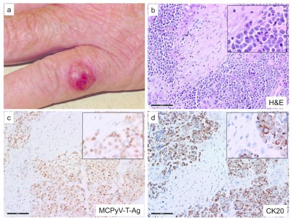
Figure 1. Clinical and microscopic images of Merkel cell carcinoma.
a) Representative MCC on the left hand of a 70-year-old man. Microscopic images with magnified insets of a primary MCC tumor with b) Hematoxylin and eosin stain showing salt and pepper chromatin pattern, frequent mitotic figures and nuclear molding characteristic of MCC; c) MCPyV Large T-Ag immunohistochemistry (CM2B4 antibody) shows viral protein expression in tumor cells but not adjacent stroma; d) Cytokeratin 20 (CK20) immunohistochemistry demonstrates characteristic dot-like peri-nuclear staining. Scale bar, 50 μM

