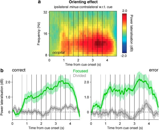
Figure 5.
Focusing of spatial attention. a, Lateralization of alpha-band activity (8–16 Hz) with respect to the cued location at lateral occipital electrodes throughout stream presentation in the focused attention condition. Gray vertical lines indicate the onsets of the premask and postmasks, and black vertical lines indicate the onsets of the eight evidence samples. b, Temporal profile of orienting-related alpha-band lateralization in the focused (green lines) and divided (gray lines) attention conditions, separately for correct choices (left) and errors (right). Conventions are the same as in a. Shaded error bars indicate SEM.

